Cellmatrix® 系列产品
细胞培养用胶原蛋白
Cellmatrix® 是拥有30年以上历史的胶原蛋白研究试剂,其中TypeⅠ-A可快速凝胶化且可调整高透明度,是非常适合用于培养胶原蛋白凝胶的产品。 除此之外,通过3D细胞培养技术还可实现接近生物体内环境的条件下进行培养。 【可应用的研究领域】细胞再生医疗研究、药学/药理学研究、三维器官、生物材料等 |
|
◆细胞培养用胶原蛋白Cellmatrix®
为配合各种用途提供以下4种类型的产品。
Cellmatrix® Type I-A ● 猪腱来源,酸可溶性的Type-I胶原蛋白 ● 浓度3.0 mg/mL、pH3的无菌溶液 ● 高凝胶强度,适合胶原蛋白・凝胶包埋培养 ● 凝胶的透明度高,容易显微镜观察 ※ 产品有效期:自生产日期起1年 |  |
Cellmatrix® Type I-P ● 猪腱来源的胃蛋白酶可溶化的Type-I胶原蛋白 ● 浓度3.0 mg/mL、pH3的无菌溶液 ● 粘性低、容易处理 ● 形成凝胶 ※ 产品有效期:自生产日期起1年 | |
Cellmatrix® Type I-C ● 猪皮来源的胃蛋白酶可溶化的Type-I胶原蛋白 ● 浓度3.0 mg/mL、 pH3的无菌溶液 ● 低粘度,适合包被 ● 几乎不会凝胶化 ※ 产品有效期:自生产日期起2年 | |
Cellmatrix® Type Ⅳ ● 用胃蛋白酶处理牛晶状体前囊精制的Type-Ⅳ胶原蛋白 ● 低粘度、适合包被 ● 浓度3.0 mg/mL、pH3的无菌溶液 ● 不会凝胶化 ※ 产品有效期:自生产日期起2年 |
◆Cellmatrix的特点
● Cellmatrix Type Ⅰ-A优化了凝胶速度和透明度,最适用于3D细胞培养的产品。
● Cellmatrix系列产品的黏度以及凝胶性能各有不同。如计划使用胶原蛋白包埋法进行培养,建议使用凝胶性能高的产品。而黏度越低的产品则越
● 容易进行处理,适合用于包被培养。下方表格整理了各产品的特点以供参考。
● Cellmatrix Type Ⅳ 不会独立形成凝胶,如想要使用此产品进行凝胶包埋培养,则需要与Cellmatrix TypeⅠ-A混合使用从而获得凝胶化的效
● 果。Cellmatrix TypeⅠ-A的含量达到40%以上时能够稳定地形成凝胶。
Cellmatrix产品 | 凝胶性能 | 包被性 |
Type Ⅰ-A | ◎ | △ |
Type Ⅰ-P | 〇 | 〇 |
Type Ⅰ-C | △ | ◎ |
Type Ⅳ | × | ◎ |

◆根据用途选择Cellmatrix® 系列

■ 胶原蛋白·凝胶包埋法(3D细胞培养)
由胶原蛋白凝胶提供的三维立体空间结构中培养细胞的方法。通过3D细胞培养技术可实现接近生物体内环境的条件下进行细胞培养。
点击此处查看实验操作

■ 胶原蛋白凝胶上培养法
细胞在胶原蛋白凝胶上培养的方法。通过在胶原蛋白凝胶上进行培养可以实现在接近生物体内环境的条件下进行细胞培养。
点击此处查看实验操作

■ 胶原蛋白包被法
将胶原蛋白分子置于已涂布的培养基质上进行细胞培养的方法。此方法在改善细胞粘附性以及通过与胶原蛋白分子接触从而激活细胞内的信号传导方面备受期待。
点击此处查看实验操作

◆胶原凝胶包埋培养套装
此套装产品中包含的实验所必要的试剂,让刚开始使用Cellmatrix®Ⅰ-A进行凝胶培养的用户也能轻松上手。 套装组成 ● Cellmatrix Type I-A ——20 mL×1 ● Ham F-12培养液——5 mL×1 ● MEM Hanks培养液——5 mL×1 ● 重构缓冲液——4mL×5 |
|
◆浓缩培养液
本产品是为了以胶原蛋白凝胶培养为目的配制的浓缩培养液。 系列产品 Ham F-12培养液(10倍浓缩) MEM Hanks培养液(10倍浓缩) DF培养液(DME:F-12=1:1)(10倍浓缩) 199培养液(10倍浓缩) DME培养液(5倍浓缩) RPMI-1640培养液(5倍浓缩) 保存条件:冷藏保存(4℃~8℃) 产品有效期:4个月 |
|
◆重构缓冲液
缓冲液组成 氢氧化钠——50 mM 碳酸氢钠——260 mM HEPES——200 mM 保存条件:冷藏保存(4℃~8℃) 产品有效期:4个月 |
|
请点击此处下载产品宣传页
Q: 请告知胶原蛋白和明胶的差异。为什么明胶在4℃做凝胶,胶原蛋白在37℃做凝胶?
A: 明胶是胶原蛋白的变性产物,具体地说,胶原蛋白是三股螺旋缠绕的结构,分子量为30万道尔顿,经热等因素而被破坏,形成无规则线圈
结构的蛋白质为明胶。所以明胶和胶原蛋白,一级结构(氨基酸组成)相同,二级结构不同。这个差异反映了胶原蛋白和明胶产生凝胶的
条件差异。
胶原蛋白保持三股螺旋缠绕的结构,在螺旋结构外侧排列着疏水性氨基酸——脯氨酸。胶原蛋白凝胶利用这个疏水性氨基酸,通过<温度
高的一方,具有高结合力>疏水结合,制备明胶。而明胶二级结构被破坏,亲水性氨基酸在外侧排列,通过氢结合和离子结合制作凝胶。
当温度高时,<氢结合力+离子结合力>克服明胶主链的热运动,不形成凝胶。但是,当变为低温时,<氢结合力+离子结合力>成为热运动
力量,明胶形成凝胶。
Q: 用明胶凝胶是否能培养?
A: 用明胶凝胶不能培养。明胶凝胶在37℃的培养条件下不会形成凝胶,将会溶化。
在组织培养液中,明胶的用途一般为:在培养皿中进行明胶涂层培养,或在明胶海绵上用福尔马林等进行桥联培养。
Q: 想购买胶原蛋白凝胶,是否有?
A: 以胶原蛋白溶液的状态作为商品销售。可根据附送的手册,进行胶原蛋白凝胶制作。
请将Cellmatrix Type I-A作为胶原蛋白凝胶培养产品,进行订购。为第一次做胶原蛋白凝胶培养法的顾客,准备了制作凝胶所必须的试剂
套装和胶原蛋白凝胶培养试剂盒。
Q: 为什么溶解于酸性溶液的胶原蛋白能进行培养?
A: 实际上,在胶原蛋白包埋细胞前,需要将酸性的胶原蛋白溶液调整成中性。
调整后的胶原蛋白因为低温保持者溶液状态,以这个状态将细胞悬液混合到胶原蛋白溶液中。
之后,将含有这个细胞的胶原蛋白溶液移到培养皿中,在37℃中加热,胶原蛋白以细胞包埋状态形成凝胶。
这个就是胶原蛋白凝胶包埋培养法的制作方法。
详细请参考在线手册。
Q: Cellmatrix有很多的胶原蛋白种类,应该选哪个才好?
A: 根据目的不同,变化使用的种类。
根据主要目的,将产品系列分成如下种类,请参考。
胶原蛋白凝胶培养试剂盒、Cellmatrix Typr I-A、Cellmatrix TypeI-P适合胶原蛋白凝胶培养法。
其他的Cellmatrix(Cellmatrix Type I-C,III,IV)不能进行胶原蛋白凝胶培养。
Cellmatrix Type I-C适合标准的涂层培养法——胶原蛋白涂层培养法。请根据客人实验的细胞及方法,考虑适合的Cellmatrix ,实验性地
考虑其他类型的胶原蛋白。例如,软骨细胞使用Cellmatrix Type II等,表皮细胞使用Cellmatrix Type IV等。
Q: 为何胶原蛋白凝胶比琼脂凝胶的培养效率好?
A: 请让我简单地说明琼脂培养法和胶原蛋白凝胶培养法的差异。
被包埋在胶原蛋白凝胶内的细胞,在胶原蛋白基质上以贴壁的状态进行增殖。 .
可是,被琼脂包埋的细胞,在琼脂基质上以未贴壁状态进行增殖。所以,贴壁依赖性细胞在胶原蛋白凝胶中进行细胞增殖,在琼脂内不进行
细胞增殖。
有报告指出:贴壁依赖性比较低的癌细胞,采用胶原蛋白凝胶培养法比采用琼脂培养法,显示高倍数的克隆形成率。
有众多报告指出:胶原蛋白凝胶法对细胞增殖、细胞分化的诱导、形态形成等有效。
关于胶原蛋白凝胶培养法的详细报告,请参考主页的论文介绍。
Q: 胶原蛋白凝胶包埋培养法和胶原蛋白、涂层培养法,有什么不同?
A: 胶原蛋白凝胶包埋培养法是在胶原蛋白凝胶内做细胞包埋,进行细胞培养的方法。
被包埋到胶原蛋白内的细胞,和体内一样进行三维增殖。
胶原蛋白涂层培养法是在培养皿上做胶原蛋白薄涂层,然后再上面进行细胞培养的方法,细胞呈单层增殖。
综上所述,胶原蛋白凝胶包埋培养法是作为细胞外基质,利用胶原蛋白的三维培养法:胶原蛋白涂层培养法是利用胶原蛋白作为细胞吸附因
子的二维培养法(单层培养法)。
Q: 想进行胶原蛋白凝胶培养,应该选哪种Cellmatrix才好?
A: 请选Cellmatrix Type I-A。还有为初学者准备的,含有制作胶原蛋白凝胶全部试剂的胶原蛋白凝胶培养试剂盒。
建议第一次使用的人购买这个胶原蛋白凝胶培养试剂盒,试剂盒货号是638-00781。
Q: 想进行胶原蛋白、涂层培养防法,应该选哪种Cellmatrix才好?
A: 请选Cellmatrix Type I-C。Cellmatrix Type I-C具有低粘度,处理容易,适合标准胶原蛋白涂层培养法。
根据细胞种类和培养条件的不同,也能得到更好的适合细胞的胶原蛋白涂层。
例如,表面细胞可使用Cellmatrix Type IV胶原蛋白等。请进行实验研讨。
Q: 想学习胶原蛋白培养法,请介绍合适的论文。
A: 详细请参考在线手册。
该手册详细地介绍了:所谓的胶原蛋白是什么、胶原蛋白培养法的效果、胶原蛋白培养发的具体方法等。
关于论文,请参考主页的论文介绍项目。
Q: 请具体地告知胶原蛋白、凝胶包埋培养方法。
A: 将作为酸性溶液的胶原蛋白溶液Cellmatrix Type I-A调整成中性。
调整后的胶原蛋白因为低温,保持着溶液状态,以这个状态将细胞悬浊液混合到胶原蛋白溶液中。之后江汉油田这个细胞的胶原蛋白溶液
移到培养皿中,在37℃中加热,胶原蛋白以细胞包埋状态形成凝胶。
关于详细 ,请参考在线手册。
Q: 请具体地告知胶原蛋白涂层的方法。
A: 将Cellmatrix Type I-C用稀盐酸(pH 3.0,约10-3 M)稀释10倍以上,放入培养皿中,在室温中静置30~60分钟。
静置后,摄取胶原蛋白溶液,以常温、无菌状态干燥培养皿。干燥后,用PBS或培养液洗涤2次,加入细胞悬液,进行普通培养。
Q: 进行胶原蛋白涂层时,向培养皿注入多少量的胶原蛋白溶液才好?
A: 胶原蛋白溶液注入量为均匀覆盖培养皿底面即可。
以1 mL/10 cm2比例为标准
Q: Cellmatrix Type IV是否能进行包埋培养?
A: 单独使用Cellmatrix Type IV胶原蛋白不能进行包埋培养。众多胶原蛋白中只有I型胶原蛋白具有凝胶化能力。
若想研究在包埋培养下的IV型胶原蛋白与细胞的互相作用,请混合I型胶原蛋白IV型胶原蛋白。Cellmatrix Type I和Cellmatrix Type IV的混
合比例,在1:2~2:1范围为好。混合比例与胶原蛋白凝胶强度的关系,请参考本公司手册<使用胶原蛋白的细胞培养法>。
Q: 所谓的胶原蛋白是不是培养基?
A: 胶原蛋白不是培养基。所谓的胶原蛋白是培养基的基质。
胶原蛋白凝胶是在培养基中作为整体结构支撑体的胶原蛋白组织,相当于生物体中的血液。
在体外的细胞作为胶原蛋白的细胞外基质存在。
将胶原蛋白作为培养基质使用的胶原蛋白凝胶包埋培养,旨在重现生物内环境的状态。
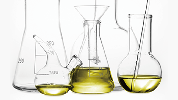
Q: 请告知胶原蛋白的稀释方法。
A: 用pH 3.0稀盐酸稀释。因为胶原蛋白具有高粘性,使用像试管一样的细小容器比较难稀释,使用三角烧瓶或离心管(例如:50 mL培养用
塑料离心管等)等粗的容器比较容易稀释。
三角烧瓶,像画圆一样摇晃10次左右:离心管,来回颠倒10次左右,进行搅拌稀释。如果,出现起泡,可静置一晚后使用,或以低速离心
分离(1500 rpm,3 min)脱气后使用。同时,开发了胶原蛋白、涂层培养用低粘度型的Cellmatrix Type I-C。可一起讨论。
Q: pH3.0盐酸是多少摩尔值?请告知制作方法。
A: pH盐酸是10-3摩尔/升。因为盐酸是强酸,浓度基本合适,调整好pH计,就可快速、正确地制作。
例如,一边用pH仪表测量,一边用巴斯德移液管一滴一滴向100 mL蒸馏水中加入1N的盐酸。
pH3.0盐酸制成后,请以高压灭菌或过滤灭菌。
| 1. | Matsunaga, M., Hatta, K., Nagafuchi, A., & Takeichi, M. (1988). Guidance of optic nerve fibres by n-cadherin adhesion molecules. Nature, 334(6177), 62-4. 【Cellmatrix® Type I-A. IF:43.07】 |
| 2. | Morishita, A. , Kumabe, S. , Nakatsuka, M. , & Iwai, Y. . (2014). A histological study of mineralised tissue formation around implants with 3d culture of hms0014 cells in cellmatrix type i-a collagen gel scaffold in vitro. Okajimas Folia Anatomica Japonica, 91(3), 57-71. 【Cellmatrix® Type I-A】 |
| 3. | Nakao, K., Itoh, M., Tomita, Y., Tomooka, Y., & Tsuji, T. (2004). Fgf-2 potently induces both proliferation and dsp expression in collagen type i gel cultures of adult incisor immature pulp cells. Biochemical & Biophysical Research Communications, 325(3), 1052-1059. 【Cellmatrix® Type I-A. IF:2.705】 |
| 4. | Udagawa, N., Takahashi, N., Akatsu, T., Tanaka, H., Sasaki, T., & Nishihara, T., et al. (1990). Origin of osteoclasts: mature monocytes and macrophages are capable of differentiating into osteoclasts under a suitable microenvironment prepared by bone marrow-derived stromal cells. Proceedings of the National Academy of Sciences of the United States of America, 87(18), 7260-7264. 【Cellmatrix® Type I-A. IF:9.58】 |
| 5. | Koide, N., Sakaguchi, K., Koide, Y., Asano, K., Kawaguchi, M., & Matsushima, H., et al. (1990). Formation of multicellular spheroids composed of adult rat hepatocytes in dishes with positively charged surfaces and under other nonadherent environments. Experimental Cell Research, 186(2), 0-235. 【Cellmatrix® Type I-A ,Cellmatrix® Type III,Cellmatrix® Type IV. IF:3.329】 |
| 6. | Jabaji, Z., Brinkley, G. J., Khalil, H. A., Sears, C. M., Nan, Y. L., & Lewis, M., et al. (2014). Type i collagen as an extracellular matrix for the in vitro growth of human small intestinal epithelium. Plos One, 9(9), e107814. 【Cellmatrix® Type I-A. IF:2.776】 |
| 7. | Shakado, S., Sakisaka, S., Noguchi, K., Yoshitake, M., Harada, M., & Mimura, Y., et al. (2010). Effects of extracellular matrices on tube formation of cultured rat hepatic sinusoidal endothelial cells. Hepatology, 22(3), 969-973. 【Cellmatrix® Type I-A,Cellmatrix® Type IV. IF:14.971】 |
| 8. | Tamura, T., Udagawa, N., Takahashi, N., Miyaura, C., Tanaka, S., & Yamada, Y., et al. (1993). Soluble interleukin-6 receptor triggers osteoclast formation by interleukin 6. Proceedings of the National Academy of Sciences of the United States of America, 90(24), 11924-11928. 【Cellmatrix® Type I-A. IF:9.58】 |
| 9. | Takahashi, N., Udagawa, N., Akatsu, T., Tanaka, H., Isogai, Y., & Suda, T. (1991). Deficiency of osteoclasts in osteopetrotic mice is due to a defect in the local microenvironment provided by osteoblastic cells*. Endocrinology, 128(4), 1792-1796. 【Cellmatrix® Type I-A. IF:3.8】 |
| 10. | Mano, H. . (1996). Mammalian mature-osteoclasts as estrogen target cells. Biochemical & Biophysical Research Communications, 223(3), 637-642. 【Cellmatrix® Type I-A. IF:2.705】 |
| 11. | Udagawa, N., Takahashi, N., Jimi, E., Matsuzaki, K., Tsurukai, T., & Itoh, K., et al. (1999). Osteoblasts/stromal cells stimulate osteoclast activation through expression of osteoclast differentiation factor/rankl but not macrophage colony-stimulating factor: receptor activator of nf-kappa b ligand. Bone, 25(5), 517-523. 【Cellmatrix® Type I-A. IF:4.36】 |
12. | Woo, J. T., Kasai, S., Stern, P. H., & Nagai, K. (2010). Compactin suppresses bone resorption by inhibiting the fusion of prefusion osteoclasts and disrupting the actin ring in osteoclasts. Journal of Bone & Mineral Research, 15(4), 650-662. 【Cellmatrix® Type I-A. IF:5.711】 |
| 13. | Ishida, K., Murofushi, M., Nakao, K., Morita, R., Ogawa, M., & Tsuji, T. (2011). The regulation of tooth morphogenesis is associated with epithelial cell proliferation and the expression of sonic hedgehog through epithelial–mesenchymal interactions. Biochemical & Biophysical Research Communications, 405(3), 455-461. 【Cellmatrix® Type I-A. IF:2.705】 |
| 14. | Nakagawa, H. , Wachi, M. , Woo, J. T. , Kato, M. , Kasai, S. , & Takahashi, F. , et al. (2002). Fenton reaction is primarily involved in a mechanism of (?)-epigallocatechin-3-gallate to induce osteoclastic cell death. Biochemical and Biophysical Research Communications, 292(1), 94-101. 【Cellmatrix® Type I-A. IF:2.705】 |
| 15. | Onodera, K. I., Fukatsu, T., Kawai, N., Yoshioka, Y., Okamoto, T., & Nakamura, H., et al. (2004). Zooxanthellactone, a novel γ-lactone-type oxylipine from dinoflagellates of symbiodinium sp.: structure, distribution, and biological activity. Bioscience Biotechnology & Biochemistry, 68(4), 848-852. 【Cellmatrix® Type I-A. IF:1.297】 |
| 16. | Torisawa, Y. S., Shiku, H., Yasukawa, T., Nishizawa, M., & Matsue, T. (2005). Multi-channel 3-d cell culture device integrated on a silicon chip for anticancer drug sensitivity test. Biomaterials, 26(14), 2165-2172. 【Cellmatrix® Type I-A. IF:10.273】 |
| 17. | Murakami, H., Takahashi, N., Tanaka, S., Nakamura, I., Udagawa, N., & Nakajo, S., et al. (1997). Tiludronate inhibits protein tyrosine phosphatase activity in osteoclasts. Bone, 20(5), 399-404. 【Cellmatrix® Type I-A. IF:4.36】 |
| 18. | Fujimoto, M., & Mihara, S. I. (1991). Two states of the l-type ca 2+ channel in pc12 cells: different sensitivity to 1,4-dihydropyridines. Neuroscience Letters, 122(1), 9-12. 【Cellmatrix® Type I-A. IF:2.173】 |
| 19. | Kawamura, S., Wakitani, S., Kimura, T., Maeda, A., Caplan, A. I., & Shino, K., et al. (1998). Articular cartilage repair: rabbit experiments with a collagen gel-biomatrix and chondrocytes cultured in it. Acta Orthop Scand, 69(1), 56-62. 【Cellmatrix® Type I-A】 |
| 20. | Tilney, L. G., Derosier, D. J., & Mulroy, M. J. (1980). The organization of actin filaments in the stereocilia of cochlear hair cells. Journal of Cell Biology, 86(1), 244-259. 【Cellmatrix® Type III,Cellmatrix® Type IV. IF:8.891】 |
| 21. | Yoshii, C., Ueda, Y., Okamoto, M., & Araki, M. (2007). Neural retinal regeneration in the anuran amphibian xenopus laevis post-metamorphosis: transdifferentiation of retinal pigmented epithelium regenerates the neural retina. Developmental Biology, 303(1), 45-56. 【Cellmatrix® Type I-C. IF:2.936】 |
| 22. | Aoki, K. , & Matsuda, M. . (2009). Visualization of small gtpase activity with fluorescence resonance energy transfer-based biosensors. NATURE PROTOCOLS, 4(11), 1623-1631. 【Cellmatrix® Type I-C. IF:11.334】 |
| 23. | Minakawa, M., Miura, Y., & Yagasaki, K. (2012). Piceatannol, a resveratrol derivative, promotes glucose uptake through glucose transporter 4 translocation to plasma membrane in l6 myocytes and suppresses blood glucose levels in type 2 diabetic model db/db mice. Biochemical & Biophysical Research Communications, 422(3), 469-475. 【Cellmatrix® Type I-C. IF:2.705】 |
| 24. | Koide, T., Homma, D. L., Asada, S., & Kitagawa, K. (2005). Self-complementary peptides for the formation of collagen-like triple helical supramolecules. Bioorganic & Medicinal Chemistry Letters, 15(23), 5230-5233. 【Cellmatrix® Type I-C. IF:2.448】 |
| 25. | Izuta, H., Shimazawa, M., Tsuruma, K., Araki, Y., Mishima, S., & Hara, H. (2009). Bee products prevent vegf-induced angiogenesis in human umbilical vein endothelial cells. Bmc Complementary & Alternative Medicine, 9(1), 45. 【Cellmatrix® Type I-C. IF:2.479】 |
| 26. | Umigai, & Naofumi. (2012). Crocetin, a carotenoid derivative, inhibits vegf-induced angiogenesis via suppression of p38 phosphorylation. Current Neurovascular Research, 9(2), 102-109. 【Cellmatrix® Type I-C. IF:1.811】 |
| 27. | Isaji, M., Miyata, H., Ajisawa, Y., Takehana, Y., & Yoshimura, N. (2010). Tranilast inhibits the proliferation, chemotaxis and tube formation of human microvascular endothelial cells in vitro and angiogenesis in vivo. British Journal of Pharmacology, 122(6), 1061-1066. 【Cellmatrix® Type I-P. IF:6.583】 |
| 28. | Tokunaga, T., Kiso, T., Namikawa, T., & Ohtsubo, Y. (1999). Camp-independent chloride secretion activated by a vasoactive intestinal peptide in a monolayer culture of human bronchial epithelial cells. Biological & Pharmaceutical Bulletin, 22(7), 745-748. 【Cellmatrix® Type I-C. IF:1.54】 |
| 29. | Aoki, Y. , Satoh, K. , Sato, K. , & Suzuki, K. T. . (1992). Induction of glutathione\r, s\r, -transferase p-form in primary cultured rat liver parenchymal cells by co-planar polychlorinated biphenyl congeners. Biochemical Journal, 281(2), 539-543. 【Cellmatrix® Type I-P. IF:4.331】 |
| 30. | Yamamoto, M., Kato, K., & Ikada, Y. (2015). Ultrastructure of the interface between cultured osteoblasts and surface-modified polymer substrates. Journal of Biomedical Materials Research, 37(1), 29-36. 【Cellmatrix® Type I-P】 |
| 31. | Ishihara, S., Haga, H. M., Mizutani, T., Kawabata, K., Shirato, H., & Nishioka, T. (2010). Integrin beta1-dependent invasive migration of irradiation-tolerant human lung adenocarcinoma cells in 3d collagen matrix. Biochemical & Biophysical Research Communications, 396(3), 651-655. 【Cellmatrix® Type I-C,Cellmatrix® Type I-P. IF:2.705】 |
| 32. | Suzuki, M., Ichikawa, K., Sakoda, A., & Sakai, Y. (1993). Long-term culture of primary rat hepatocytes with high albumin secretion using membrane-supported collagen sandwich. Cytotechnology, 11(3), 213. 【Cellmatrix® Type I-P. IF:1.672】 |
| 33. | Nakaoka, R. , Tsuchiya, T. , Kato, K. , Ikada, Y. , & Nakamura, A. . (1997). Studies on tumor-promoting activity of polyethylene: inhibitory activity of metabolic cooperation on polyethylene surfaces is markedly decreased by surface modification with collagen but not with rgds peptide. Journal of biomedical materials research, 35(3), 391-397. 【Cellmatrix® Type I-P】 |
| 产品编号 | 产品名称 | 产品规格 | 产品等级 | 备注 |
| 631-00651 | Cellmatrix Type I -A (Collagen, Type I, 3 mg/mL, pH 3.0) 猪腱来源酸可溶性Type I-A骨胶原,高凝胶强度 (3 mg/mL) |
20 mL | - | - |
| 637-00653 | Cellmatrix Type I -A (Collagen, Type I, 3 mg/mL, pH 3.0) 猪腱来源酸可溶性Type I-A骨胶原,高凝胶强度 (3 mg/mL) |
100 mL | - | - |
| 638-00661 | Cellmatrix Type I -P (Collagen, Type I, 3 mg/mL, pH 3.0) 猪腱来源酸可溶性Type I-P骨胶原,高凝胶强度 (3 mg/mL) |
20 mL | - | - |
| 634-00663 | Cellmatrix Type I -P (Collagen, Type I, 3 mg/mL, pH 3.0) 猪腱来源酸可溶性Type I-P骨胶原,高凝胶强度 (3 mg/mL) |
100 mL | - | -- |
| 631-00771 | Cellmatrix Type I -C(Collagen, Type I, 3 mg/mL, pH 3.0) 猪皮来源胃蛋白酶可溶Type I-C,低黏度 (3 mg/mL) |
20 mL | - | - |
| 637-00773 | Cellmatrix Type I -C(Collagen, Type I, 3 mg/mL, pH 3.0) 猪皮来源胃蛋白酶可溶Type I-C,低黏度 (3 mg/mL) |
100 mL | - | - |
| 638-05921 | Cellmatrix Type IV(Collagen, Type IV, 3 mg/mL, pH 3.0) 澳大利亚和新西兰产牛水晶体前包来源胃蛋白酶可溶化Type IV 骨胶原,低黏度 (3 mg/mL) |
5 mL | - | - |
| 634-05923 | Cellmatrix Type IV(Collagen, Type IV, 3 mg/mL, pH 3.0) 澳大利亚和新西兰产牛水晶体前包来源胃蛋白酶可溶化Type IV 骨胶原,低黏度 (3 mg/mL) |
20 mL | - | - |
| 632-05924 | Cellmatrix Type IV(Collagen, Type IV, 3 mg/mL, pH 3.0) 澳大利亚和新西兰产牛水晶体前包来源胃蛋白酶可溶化Type IV 骨胶原,低黏度 (3 mg/mL) |
100 mL | - | - |
| 产品编号 | 产品名称 | 产品规格 | 产品等级 | 备注 |
| 638-00781 | Collagen Gel Culturing Kit 胶原凝胶包埋培养套装 |
1 kit | - | - |
| 产品编号 | 产品名称 | 产品规格 | 产品等级 | 备注 |
| 630-29661 | Concentrated culture solution Ham's F-12 culture solution 浓缩培养液 Ham F-12培养液 |
100 mL | - | - |
| 636-29641 | Concentrated culture solution MEM-Hank's culture solution 浓缩培养液 MEM Hanks培养液 |
100 mL | - | - |
| 632-29621 | Concentrated culture solution DF culture solution 浓缩培养液 DF培养液 |
100 mL | - | - |
| 635-29611 | Concentrated culture solution 199 culture solution 浓缩培养液 199培养液 |
100 mL | - | - |
| 639-29631 | Concentrated culture solution DME culture solution 浓缩培养液 DME培养液 |
100 mL | - | - |
| 633-29651 | Concentrated culture solution RPMI-1640 culture solution 浓缩培养液 RPMI-1640培养液 |
100 mL | - | - |
| 产品编号 | 产品名称 | 产品规格 | 产品等级 | 备注 |
| 635-00791 | Reconstitution Buffer 溶解用缓冲液(胶原蛋白/凝胶培养用) |
4 mL×15 | - | - |
| 免责声明 |
|
1. 本公司密切关注本网站发布的内容,但不保证发布内容的准确性、完整性、可靠性和最新性等。 2. 本公司不保证使用本网站期间不会出现故障或计算机病毒污染的风险。 3. 无论何种原因,使用本网站时给用户或第三方造成的任何不利或损害,本公司概不负责。此外,对于用户与其他用户或第三方之间因本网站发生的任何交易、通讯 3. 或纠纷,本公司概不负责。 4. 本网站可提供的所有产品和服务均不得用于人体或动物的临床诊断或治疗,仅可用于科研等非医疗目的。如任何用户将本网站提供的产品和服务用于临床诊断或治 4. 疗,以及其他特定的用途或行为,本公司概不保证其安全性和有效性,并且不负任何相关的法律责任。 |